Diabetic retinopathy: How diabetes affects the eyes
Scientific support: Prof. Dr. Rainer Guthoff, Dr. Gidon Bönhof
Diabetes can damage the smallest blood vessels in the eyes and thus also the retina. Many people with diabetes do not initially notice these changes but as time passes problems may develop, from slight visual impairment through to blindness.
There are many types of eye diseases caused by diabetes: As well as changes to the retinal vessels (retinopathy), and the macula (maculopathy), inflammation of the upper and lower lid and cataracts have been observed. The macula is the area on the retina responsible for the sharpest vision and is where most of the cells responsible for sight are located, including those for color perception and the ability to read. There may also be changes to the pressure inside the eye and effects on all nerves involved in the function of the eye. Changes to the pressure inside the eye can also lead to glaucoma.

Contents
- What increases the risk of diabetic eye disease?
- How is diabetic retinopathy diagnosed?
- How does retinopathy develop in people with diabetes?
- How can eye diseases associated with diabetes be prevented?
- What does the ophthalmologist examine?
- How is diabetic retinopathy treated?
- How common are diabetes-related eye diseases?
- Help for people with diabetes and visual impairments
1. What increases the risk of diabetic eye disease?
The most well-established risk factors for developing diabetic eye disease in people with diabetes are:
- Smoking
- Persistently elevated blood glucose levels
- Elevated blood pressure
- Fat metabolism disorders
- Long-term diabetes
- Kidney damage due to diabetes (diabetic nephropathy)
2. How is diabetic retinopathy diagnosed?
Many of those affected do no initially notice that they have diabetic eye disease. For this reason people with diabetes should visit an ophthalmologist at least every two years to check for changes to the retina if no further risk factors are present. If they have one or more risk factors for developing diabetic eye disease, they should be examined every year.
The German Diabetes Association (DDG) recommends that people with type 1 diabetes begin these check-ups of the ocular fundus from the age of 11 or at least 5 years after the initial diagnosis. For people with type 2 diabetes, these check-ups should begin immediately after the initial diagnosis.
It is only usually in the advanced stages that patients notice diabetic retinal damage.
Examples of the symptoms of diabetic retinal damage are:
- blurred vision
- unclear vision
- dark patches or a "red blur" in the visual field
Frequent small bleeds or poor blood supply to the tissue can damage the retina so badly that it detaches. People notice they have retinal detachment by flashes of light or a "sooty rain”. If the part of the retina that has the sharpest vision (the macula) is affected, they notice a dark curtain moving across their vision. Retinal detachment and damage to the macula can progress to complete blindness.
3. How does retinopathy develop in people with diabetes?
Elevated blood glucose levels mean that the fat and proteins are deposited in the smallest and small blood vessels, causing the walls to thicken. This leads to localized outpouchings called microaneurysms. They may leak blood, which ophthalmologists and opticians can see as tiny dots on the retina. Because the fluid is leaking, the retina swells. Fats from blood plasma can be deposited. This is evident in an examination of the fundus of the eye, where they appear as “hard exudate”. In this initial stage no new blood vessels are formed. Medical specialists refer to this as non-proliferative retinopathy.
Over time because the blood supply to the vessels is consistently poor, the retina is not supplied with oxygen and its state deteriorates further. New blood vessels are formed to counteract this. This proliferative form of diabetic retinopathy causes blood vessels that have grown into the vitreous body to burst. The vitreous body is the clear gel that fills the space between the lens and the retina of the eyeball. Therefore, those affected suddenly has blurred vision. In a small proportion of patients with diabetes and after a longer phase of diabetes, this can lead to blindness.
Both types of diabetic retinopathy can be accompanied by maculopathy (fluid collection in the region of the macula). Depending on the severity, the function of the sensory cells at the point of sharpest vision in the retina is restricted to varying degrees. If left untreated, maculopathy can lead to blindness and is the most common cause in people with diabetes.
Good to know:
There are two types of diabetic retinopathy: non-proliferative retinopathy and proliferative retinopathy. Proliferative retinopathy causes an unnatural development of new blood vessels. Both forms can result in diabetic maculopathy. Unfortunately, these changes cause an average of 1 in 5000 people with diabetes to go blind every year.
4. How can eye diseases associated with diabetes be prevented?
It is essential to attend scheduled appointments with an ophthalmologist. Damage to the retina and other complications can be reduced or even prevented through optimal long-term management of blood sugar and blood pressure. A healthy lifestyle can also help prevent complications of diabetes.
This includes:
- Stopping smoking (smoking impairs blood circulation, including in the eyes)
- Being active (physical activity promotes blood circulation in the eyes and has anti-inflammatory effects)
- A balanced and healthy diet (so that the eyes receive sufficient vitamins and nutrients)
Good to know:
Quitting smoking, getting enough exercise, and eating a healthy diet also have positive effects on the risk factors high blood pressure and fat metabolism disorders.
5. What does the ophthalmologist examine?
Generally, the examination begins with some questions, for example, on medication use or any other preexisting conditions.
To examine the ocular fundus, the medical team uses eye drops to dilate the pupil. This allows for better assessment of the retina. In some cases, the blood circulation in the ocular fundus is examined using a special camera. For this, the ophthalmologist will first inject a coloring agent into the vein.
The ophthalmologist uses a magnifying glass that reflects the ocular fundus and the macula under a slit lamp, a special type of microscope. Visual acuity is then tested using symbols on a reading chart, which the person undergoing testing reads aloud from a set distance. Another type of microscope can then be used to examine the anterior segment of the eye.
6. How is diabetic retinopathy treated?
Doctors treat people with diabetic eye disease by strictly managing their blood glucose and blood pressure. Other risk factors such as smoking should also be avoided to prevent loss of vision and blindness. If the diabetic retinopathy advances, laser treatment may be carried out which allows the tissue to grow together tighter to prevent retinal detachment.
Laser treatment of the retina (laser coagulation) cannot always be used in cases of advanced cataracts or scar tissue formation in the vitreous body. It is only used in cases that are not too advanced. If there are already severe complications such as hemorrhaging in the vitreous body or retinal detachment, the eyes can be saved from complete blindness by surgically removing the vitreous body.
A more recent treatment option for maculopathy involves injecting steroids or vascular endothelial growth factor inhibitors (VEGF inhibitors) directly into the vitreous body space. They block the growth factors responsible for the development of new, leaking blood vessels. These drugs then inhibit further growth of the vessels. In studies, VEGF inhibitor injections have been shown to reverse even severe forms of retinopathy and reduce the number of cases of diabetes-related blindness.
7. How common are diabetes-related eye diseases?
About a quarter of people with type 1 diabetes (25 out of every 100 people with type 1 diabetes) will develop diabetic retinopathy at some point in their life. From puberty, retinopathy in children with type 1 diabetes is rare. In people with type 2 diabetes, the incidence is about half as high at 12.5 percent (13 out of every 100 people with type 2 diabetes). One in three already have some degree of retinal changes when they are diagnosed with type 2 diabetes. The further progression of this diabetes-associated eye disease depends on the management of blood sugar and blood pressure levels.
Good to know:
Diabetic retinopathy is the most common cause of blindness in adults aged 20 to 74 in developed countries.
8. Help for people with diabetes and visual impairments
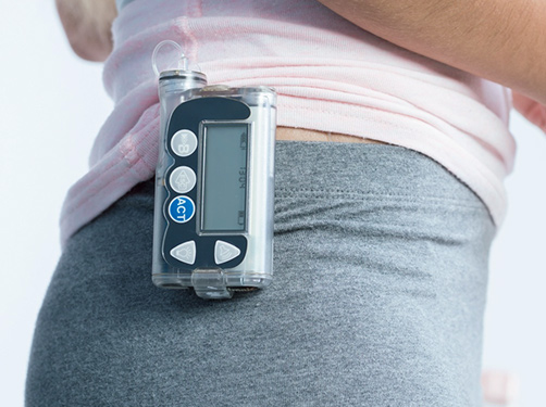
It can be difficult for people with severe visual impairments or those who are blind to independently manage their diabetes therapy. Some form of visual acuity is usually needed to read and enter information when using insulin pumps, blood sugar measurement devices, or continuous glucose monitors (CGM systems). However, technologies and aids for the treatment of diabetes are often not designed to be suitable for all.
There are very few medical devices available that those with severe visual impairment or blindness can use independently.
For example, blood sugar measurement devices with a large high-contrast and backlit display are very helpful. There are now blood sugar measurement devices that have sound or speech output or that can be connected to a smartphone app that reads out the results. There are also insulin pumps that use sounds to make it possible to differentiate between different alarms. Some types of pumps can be read and programmed using a PC or controlled using a smartphone. For work on PCs, there are screen reading programs available for visually impaired and blind people.
Good to know:
The German Federation of the Blind and Partially Sighted (DBSV; www.dbsv.org) provides advice to people with diabetes and visual impairments and advocates for needs-based care using medical technical devices. The advice and support offered by the DBSV can be found at https://blickpunkt-auge.de (Websites in German).
Sources:
American Diabetes Association: Microvascular Complications and Foot Care: Standards of Medical Care in Diabetes – 2021. In: Diabetes Care, 2021, 44: S151-S167
Bryl, A. et al.: The Effect of Diet and Lifestyle on the Course of Diabetic Retinopathy-A Review of the Literature. In: Nutrients, 2022, 14: 1252
Bundesärztekammer et al.: Patientenleitlinie zur Nationalen Versorgungsleitlinie Diabetes – Schäden an der Netzhaut: Vorbeugen und behandeln. 2. Auflage. Version 2. 2016
Bundesärztekammer et al.: Nationale Versorgungsleitlinie Prävention und Therapie von Netzhautkomplikationen bei Diabetes. Langfassung. Version 2. 2015
Patienten-Information.de: Diabetes – Schäden an der Netzhaut des Auges. (Letzter Abruf: 30.12.2022)
Raman, R. et al.: A Paradigm Shift in the Management Approaches of Proliferative Diabetic Retinopathy: Role of Anti-VEGF Therapy. In: Clin Ophthalmol, 2022, 16: 3005-3017
Scanlon, P. H.: Improving the screening of risk factors in diabetic retinopathy. In: Expert Rev Endocrinol Metab, 2021, 17: 235-243
Solomon, D. et al.: Diabetic Retinopathy: A Position Statement by the American Diabetes Association. In: Diabetes Care, 2017, 40: 412-418
Trott, M. et al.: Associations between diabetic retinopathy and modifiable risk factors: An umbrella review of meta-analyses. In: Diabet Med, 2022, 39: e14796
As of: 30.12.2022